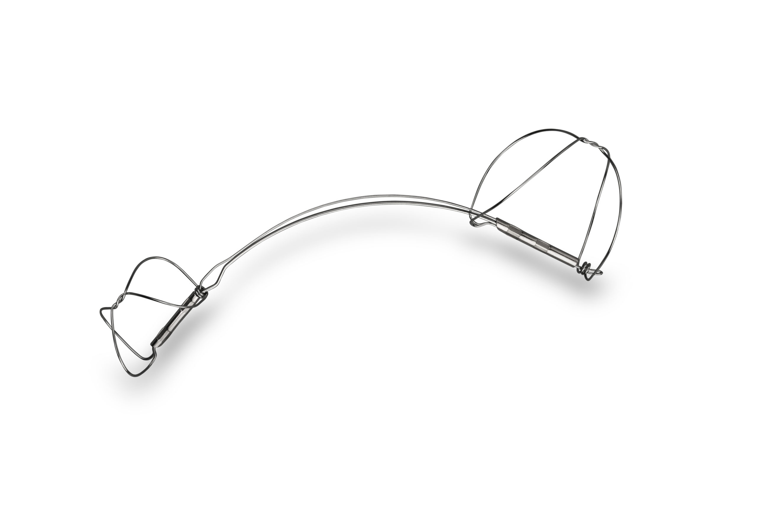
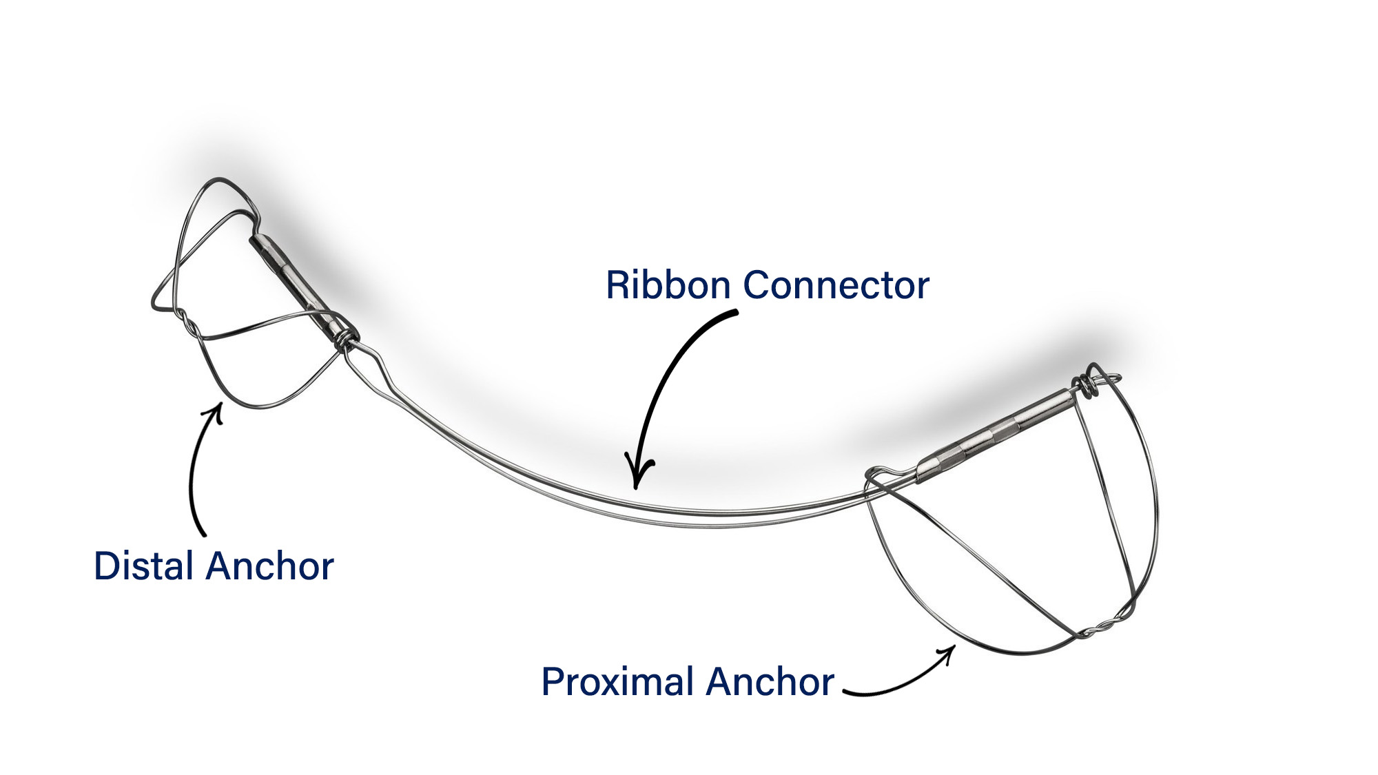
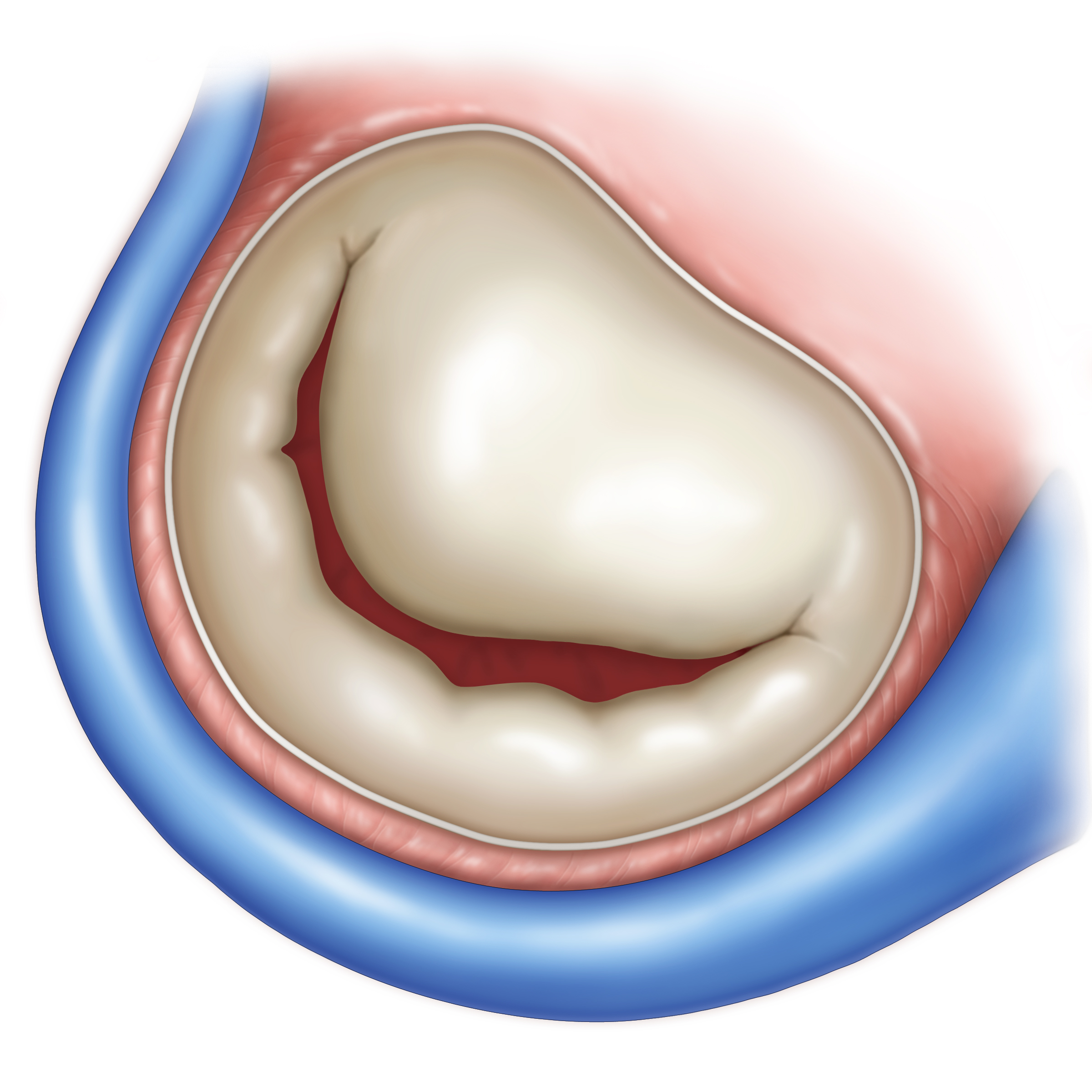
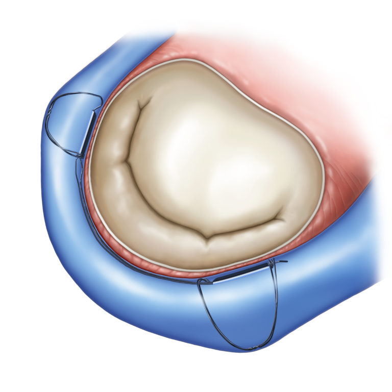
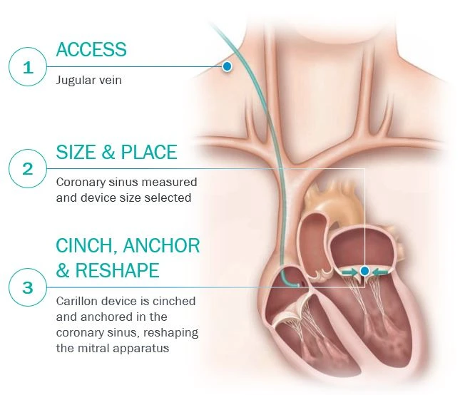
Two anchors joined by a shaping ribbon are positioned in the coronary sinus and great cardiac vein. Tension applied to the implant reduces the mitral annulus to improve leaflet coaptation, without clipping or replacing the leaflets.
Before final release, the operator assesses implant position, coronary arterial flow and the effect on mitral regurgitation. The implant can be recaptured while it remains connected to the handle assembly.
Before implantation · original illustrationWith the CARILLON implant · original illustration
Venous access and delivery catheter
CLINICAL EVIDENCE
The CARILLON clinical programme.
Nine study summaries, separating randomised evidence, prospective studies, registries and long-term follow-up. Updated 19 October 2026.
2009 · Prospective
AMADEUS
Design
Prospective multicentre, single-arm study.
Population
48 enrolled; 30 permanent implants.
Follow-up
6 months.
Findings
Feasibility of indirect annuloplasty; assessment of mitral regurgitation, functional capacity and safety.
Interpretation
Early uncontrolled study using a first-generation device.
Schofer J et al. Circulation. 2009;120:326–333. DOI: 10.1161/CIRCULATIONAHA.109.849885.
Regurgitant volume: −7.1 vs +3.3 mL/beat (p=0.049). LVEDV: −10.4 vs +6.5 mL (p=0.03).
Interpretation
Proof-of-concept echo endpoint. Paired regurgitant-volume data available in 68/120 patients; no demonstrated mortality benefit.
Witte KK et al. JACC: Heart Failure. 2019;7:945–955. DOI: 10.1016/j.jchf.2019.06.011.
2020 · Long-term follow-up
Pooled follow-up
Design
Pooled analysis of prospective studies.
Population
74 treated patients with long-term vital status available.
Follow-up
Up to 5 years.
Findings
Kaplan–Meier survival: 83.6% at 1 year, 67.9% at 3 years and 56.2% at 5 years.
Interpretation
Selected cohort without a long-term randomised control; prognostic associations do not establish causation.
Lipiecki J et al. Cardiovascular Revascularization Medicine. 2020;21:712–716. DOI: 10.1016/j.carrev.2020.02.012.
2024 · Registries / real world
CINCH · first cohort
Design
Postmarket single-arm registry.
Population
101 patients at 13 German centres.
Follow-up
Up to 5 years.
Findings
All-cause mortality: 40.1%; heart-failure hospitalisation: 53.9%; composite: 66.4%. NYHA and MR-grade improvements persisted during follow-up.
Interpretation
No comparator; early cohort overlaps the later 228-patient registry and must not be added to it.
Yildiz M et al. Cardiovascular Revascularization Medicine. 2024;60:35–40. DOI: 10.1016/j.carrev.2023.09.007.
2025 · Registries / real world
CINCH · update
Design
Prospective multicentre registry.
Population
228 patients at 22 centres; 51% with LVEF ≥50%.
Follow-up
Median 2 years; range 0–5 years.
Findings
At 1 year: NYHA III/IV fell from 81% to 29%; MR 3+/4+ from 84% to 9%; serious device/procedure-related events: 1.8%.
Interpretation
5-year estimates: mortality 46.9%, HF hospitalisation 54.0%. Uncontrolled registry; not every patient had 5-year follow-up.
Fichtlscherer S et al. Structural Heart. 2025;9:100738. DOI: 10.1016/j.shj.2025.100738.
2025 · Registries / real world
Real-world experience
Design
Retrospective single-centre study.
Population
204 consecutive patients planned for implantation; age 83 ±6 years.
Follow-up
3 months in successfully implanted patients.
Findings
NYHA improved ≥1 class in 94%; MR decreased ≥1 grade in 91% of assessed patients.
Interpretation
Uncontrolled before/after data from successfully implanted, evaluable patients, not all referred patients.
Priebe-Brämer H et al. ESC Heart Failure. 2025;12:4410–4418. DOI: 10.1002/ehf2.70016.
2026 · Long-term follow-up
REDUCE FMR · 10-year follow-up
Design
Single-centre observational extension.
Population
17 treated REDUCE FMR patients.
Follow-up
Up to 10 years.
Findings
10-year overall survival: 35%; cardiovascular survival: 66%. Median survival: 5.6 years.
Interpretation
Overall and cardiovascular survival are different endpoints. Very small cohort without long-term control; exploratory results.
Lipiecki J, Kałmucki P, Miller LE, Durel N. Global Cardiology. 2026;4. DOI: 10.4081/cardio.2026.106.
These studies address different populations, generations of device and endpoints. Cohorts overlap in pooled and registry reports; patient numbers must not be summed. Observational survival estimates do not establish superiority to other therapies. Treatment decisions follow the applicable IFU and Heart Team assessment.
Further publications
Khan MS et al. Functional outcomes with Carillon device over 1 year in patients with functional mitral regurgitation of Grades 2+ to 4+: results from the REDUCE-FMR trial. ESC Heart Failure. 2021. DOI: 10.1002/ehf2.13273.
Khan MS et al. Effect of Carillon Mitral Contour System on patient-reported outcomes in functional mitral regurgitation: an individual participant data meta-analysis. ESC Heart Failure. 2021. DOI: 10.1002/ehf2.13301.
Anker SD et al. Percutaneous Mitral Valve Annuloplasty in Patients With Secondary Mitral Regurgitation and Severe Left Ventricular Enlargement. JACC Heart Failure. 2021. DOI: 10.1016/j.jchf.2021.03.002.
Giallauria F et al. Individual patient data meta-analysis of the effects of the CARILLON mitral contour system. ESC Heart Failure. 2020;7:3383–3391.
CARILLON / VIDEOS & CASES
The procedure, step by step.
Original procedure videos, followed by frames from a clinical fluoroscopic sequence. Select a video to play it here.
A closer look at a clinical case.
Three original frames from the same patient sequence, at 00:05, 00:20 and 00:38.
Source: Cardiac Dimensions media kit. Experimental video: LifeTec Group credit retained in the original. Clinical images show one illustrative case and do not establish treatment outcomes.
Patient selection and product information.
The June 2026 European IFU indicates CARILLON for symptomatic secondary (functional) mitral regurgitation despite guideline-directed medical therapy (NYHA ≥2), when annular dilatation is the primary contributor.
Contraindications include previous mitral valve replacement or an annuloplasty ring, and existing devices in the coronary sinus/great cardiac vein except CRT leads implanted for at least six months. Consult the complete IFU for warnings, precautions, risks, sizing and implantation instructions.
Original European IFU · Revision AN · 17 June 2026.
MR Conditional.
Non-clinical testing supports scanning only under all of the following conditions:
Static magnetic field: 1.5 T or 3 T.
Maximum spatial field gradient: 1,900 gauss/cm (19 T/m).
Whole-body averaged SAR: ≤2 W/kg, Normal Operating Mode.
Under these conditions, the expected maximum temperature rise is less than 3.2°C after 15 minutes of continuous scanning. At 3 T with a gradient-echo sequence, the image artefact extends approximately 20 mm from the implant.
Source: European IFU LAB-1505-01AN, 17 June 2026, section XI. Refer to the complete IFU before an MRI examination.
RESOURCES
Documents and insights.
Direct access to resources for each device family.